Sentinel Node mapping or lymphoscintigraphy is a procedure performed in the Nuclear Medicine department to help the physician localize and remove the sentinel lymph node of a tumour. Lymph nodes are found throughout your body and act as collection points and filters for waste products, fluids and other cellular debris.
A tumour’s sentinel node is the lymph node that drains the area containing the tumour and is one of the mechanisms that allow cancer cells to spread. A lymphoscintigraphy procedure will help the surgeon to locate, identify and remove the sentinel node while leaving other unrelated lymph nodes in the area.
Preparation
As preparation for sentinel node mapping, it is highly recommended that you treat the site to be injected with a topical anesthetic.
For Melanoma, the site to be injected is typically where the biopsy was done and there should be a small scar marking it.
For Breast Cancer, the peri-areolar area is typically the site to be injected.
PLEASE INFORM YOUR DOCTOR OR THE TECHNOLOGIST IF YOU
ARE, OR THINK YOU MIGHT BE, PREGNANT OR BREASTFEEDING.
Procedure
Check-in at the Diagnostic Imaging Department at the Sault Area Hospital for your appointment.
Sentinel node mapping is performed by giving 4 injections of a radioactive tracer (99-Technesium Sulphur Colloid) around the biopsy scar (melanoma) or peri-areolar region (breast cancer). The injections are given just below the surface of the skin and will sting unless the area has been previously treated with a topical anesthetic (see preparation instructions)
For melanoma, images are acquired immediately following injection with delayed images acquired 1 to 2 hours after injection. The delayed images will consist of a whole-body scan (head to toe) and a SPECT/CT (Single Photon Emission Computerized Tomography) around the sentinel node. To perform a SPECT image the camera will slowly rotate around your body taking pictures from various angles for about 10-12 min. Following that, a low dose CT scan will be acquired and the images will be fused together allowing the physician to get an accurate location of the sentinel node.
Additionally, the technologist may place a mark on your skin using a permanent marker directly above the sentinel node. This mark may help the surgeon know exactly where the sentinel node is located for the operation.
For breast cancer, images will be acquired 1 to 2 hours after injection. A series of images will be acquired from various angles around the injection site to help locate the sentinel node.
Following your test, there are no restrictions and you will be able to drive as you normally would. If you are planning on going into the United States of America you will set off the radiation detectors for approximately 2-3 days. We can provide a letter explaining why you are setting off the detectors however you will still be stopped.
Once your scan is complete it will be examined by a radiologist and a report will be sent to the ordering doctor. Follow up your test with the ordering physician or your family doctor.
The Injection
The amount of radioactive tracer injected for Sentinel Node Mapping scan is small enough that there are no additional requirements or precautions to protect others from radiation exposure. The radiation exposure to you is less than a standard x-ray. Most of the injection will be cleared out of your body after 24 hours.
Allergic reactions to the material are extremely rare, and you should not notice any changes from the material injected.
To get the injection to localize in the sentinel node, it is injected just below the surface of the skin and is filtered out by the lymphatic system. The Lymph node that drains the area injected will collect the radioactive injection as your body works to remove it from your system.
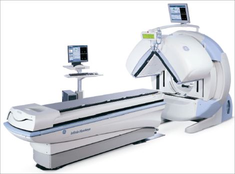
The Camera
For sentinel node mapping we acquire the images using a gamma camera. Our cameras do not emit any radiation unless performing a SPECT/CT and are not a hazard to anyone in the vicinity while acquiring. Additionally, our cameras do not make any loud noises. The cameras we use have 2 detectors: one above you and one below you. Dual camera heads allow two views to be acquired at the same time, and for SPECT imaging to be acquired in half the time.