The Myocardial Perfusion scan is a form of stress test performed in Nuclear Medicine to evaluate the blood supply to the muscle tissue of the heart (Myocardium). With this test we are not looking at how blood travels through the heart, but rather, we are looking at whether or not the muscle tissue (heart wall) is getting the blood and supplies it requires to pump properly.
The test involves two sets of images: blood supply while under stress and blood supply while at rest. At the Sault Area Hospital, we employ a two-day protocol, one day for stress, one day for rest.
Some of the main reasons why your doctor may order this test are:
- Chest pain not typically associated with a specific pathology
- Rule out reversible Ischemia (lack of blood under stress; sufficient blood at rest) Shortness of Breath
- Preoperative workup for surgery
- Unable to perform an exercise stress test
- Follow up from other testing
Preparation
For a myocardial perfusion scan, you are required to be fasting for 4 hours prior to your appointment and no caffeine (coffee, tea, pop, chocolate, etc.) for 12 hours prior to your appointment.
If you are performing the exercise stress portion of the test you may be required to be off of beta-blocker medication (Metoprolol, Atenolol, etc.) for 48 prior to your appointment.
You are able to take your normal medications with a small amount of water on the morning of the test unless otherwise told not to.
Please bring a list of your medications with you.
PLEASE INFORM YOUR DOCTOR OR THE TECHNOLOGIST IF YOU
ARE, OR THINK YOU MIGHT BE, PREGNANT OR BREASTFEEDING.
Procedure
Check-in at the Diagnostic Imaging Department at the Sault Area Hospital for your appointment.
The Sault Area Hospital uses a two-day protocol for the myocardial perfusion scan, which means you are required to attend two days in a row. The reason for two days is to give time for the tracer to clear out of the heart between each day.
On the first day, your heart will be examined either under stress or at rest and on the second day your heart will be examined under the other condition.
To examine your heart at rest you will be given a small injection of a radioactive tracer (Tc-99 Sestamibi). You should not feel anything from the radioactive tracer and reactions are extremely rare. After injection of the tracer, you will be able to have something to eat and drink if you like. You will be required to return to the nuclear medicine waiting room approximately 30-40 minutes after injection for imaging.
Examining your heart under stress can be carried out using one of two different methods: walking at various speeds on a treadmill or by using a pharmacologic agent to induce a stress-like response within the heart. The method of stress for your test will be determined by the ordering physician to match your current medical condition. Both methods are equally valid and produce similar results.
The treadmill method for examining your heart under stress is very similar to having an exercise stress test. You will be set up on a 12-lead electrocardiogram (ECG) to monitor your heart activity throughout the test. An intravenous line (IV) will be inserted to allow for venous access during the test. You will begin on the treadmill at a light walking pace and every three minutes the treadmill will increase slightly in speed and elevation. You will continue on the treadmill until you reach 85% of your maximum predicted heart rate (220-your age). Once you reach 85% of your target heart rate you will be given a small injection of a radioactive tracer (Tc-99 Sestamibi) through the IV line and asked to continue on the treadmill for another minute to complete the protocol. Once the protocol is completed you will be taken off the treadmill and monitored until your heart rate and blood pressure have returned to normal levels. After you have been disconnected you will be able to have a light snack and are required to return to the nuclear medicine department for imaging.
The pharmacological method for examining the heart under stress is good for anyone who might have difficulties walking on a treadmill at various speeds. Before performing the pharmacological stressing you will be connected to a 12-lead ECG and your resting blood pressure will be taken. An intravenous line (IV) will be set up to administer the drugs for the test. To perform the test you will be slowly administered a dose of Persantine (Dipyridamole) over the course of 4 minutes. Persantine will act as a vasodilator and dilate blood vessels throughout your body. Your heart rate typically increases a little bit; we ARE NOT trying to increase your heart and it is very rare to feel an increased heart rate.
Some of the more common side effects of Persantine include; chest discomfort, dizziness, flushing, and headaches. Four minutes after the Persantine has been administered you will be given a very small dose of the radioactive tracer (Tc-99 Sestamibi) through the IV line. Approximately 2 minutes after the radioactive tracer you will be administered Aminophylline to reverse any symptoms you might be feeling from the Persantine. After the Aminophylline you should be feeling similar to how you felt before the test and can have something to eat at this time. Diabetic patients might experience a slightly lowered blood sugar level and are recommended to bring a hard candy or juice for after the test. About an hour following the test you will be required to return to the nuclear medicine department for imaging.
Following your test, there are no restrictions and you will be able to drive as you normally would. If you are planning on going into the United States of America you will set off the radiation detectors for approximately 2-3 days. We can provide a letter explaining why you are setting off the detectors however you will still be stopped.
Once your scan is complete it will be examined by a cardiologist and a report will be sent to the ordering doctor within a week. Follow up your test with the ordering physician or your family doctor.
The Injections
The amount of radioactive tracer injected for a Myocardial Perfusion Scan is small enough that there are no additional requirements or precautions to protect others from radiation exposure. The radiation exposure to you from each injection is a little more than a standard chest x-ray and less than half of that from a CT scan. Most of the injection will be cleared out of your body after 24 hours.
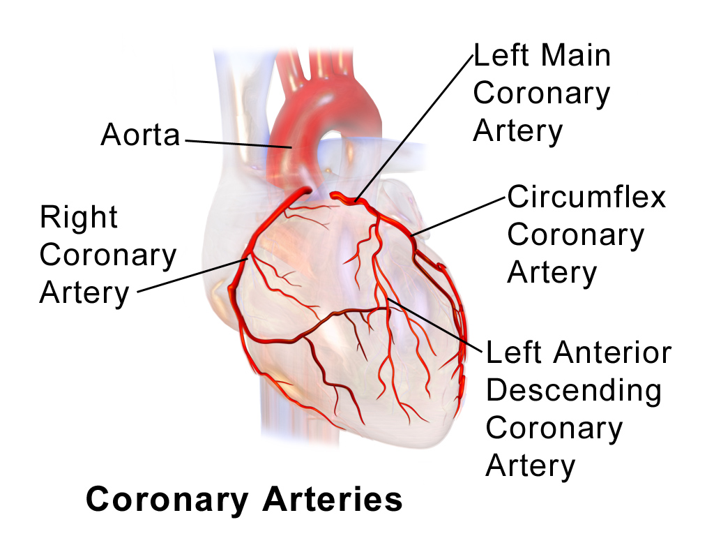
The radioactive injection used for this test is designed to be absorbed by the myocardium (heart muscle) in proportion to the amount of blood supplied to the area. This allows for the evaluation of blood supply to the heart muscle tissue (heart wall). If you have a blockage in your coronary arteries it will decrease the blood flow downstream of the blockage resulting in less absorption of the radioactive material by the muscle tissue in the affected area. The images will show exactly where the radioactive injection went within the heart.
Allergic reactions to the materials used are extremely rare. You should not feel anything or notice any changes from the radioactive material injected. If you are receiving an injection of Persantine some of the more common side effects are: chest discomfort, dizziness, flushing, and headaches.
Imaging
There will be two sets of images acquired during this procedure; one on the first day and one on the second day. To acquire the images you will be laying on your back connected to a three-lead electrocardiogram (ECG). One lead will be placed by each shoulder and the third lead will be placed just below the ribs on your left side. The ECG will allow the camera to synchronize image acquisition with your heart rate; taking a rapid series of pictures each time your heart beats. The camera will be in an upside-down V (Λ) configuration and come in very close to your chest. Your head does not usually go under the camera. Every 25 seconds the camera will rotate a couple of degrees counter-clockwise, moving around to your left side. Each set of images will take approximately 15 minutes to acquire.
The Camera
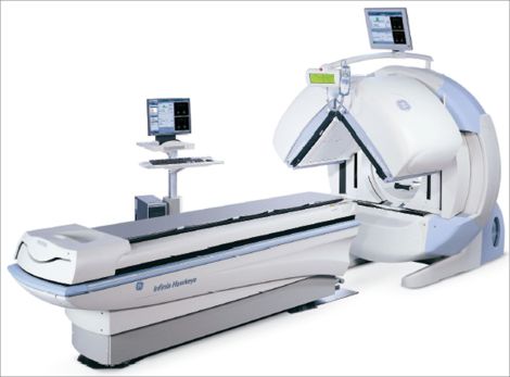
For a Myocardial Perfusion Scan, we acquire the images using a gamma camera. A gamma camera is a type of radiation detector that is used to show us how the radioactive tracer is distributed throughout your myocardium. Our cameras do not emit radiation or loud noises and are not a hazard to anyone in the vicinity while acquiring. The cameras we use have 2 detectors that will be 90° to each other (see image below) allowing for faster image acquisition.