A MUGA (Multi Gated Acquisition) Scan is a Nuclear Medicine procedure performed to evaluate the functioning of the hearts’ left Ventricle. The left ventricle is one of the four heart chambers and is responsible for pumping blood throughout the body. One of the main ways to evaluate the left ventricle functioning is to calculate an ejection fraction.
The ejection fraction is the amount of blood pushed out when the heart contracts (systolic) compared to the amount of blood when the ventricle is full (diastolic).
Your doctor may order a MUGA scan to evaluate:
- Possible decreased left ventricle functioning
- Ensure a good ejection fraction before, during and after some chemotherapies that are toxic to the heart
- Follow up on decreased or borderline results from previous tests
Preparation
There is no preparation for a MUGA scan.
PLEASE INFORM YOUR DOCTOR OR THE TECHNOLOGIST IF YOU
ARE, OR THINK YOU MIGHT BE, PREGNANT OR BREASTFEEDING.
Procedure
Check-in at the Diagnostic Imaging department at the Sault Area Hospital for your appointment.
To perform a MUGA scan we are required to label your red blood cells with a radioactive tracer. To label your red blood cells we begin by injecting a labelling agent (Gluceptate) into a vein. Gluceptate is a drug that is designed to be taken up by your red blood cells and facilitates the binding of the radioactive tracer to the red blood cells. You shouldn’t feel anything from the Gluceptate injection. Once injected, you will be asked to wait between 10 and 15 minutes giving time for the Gluceptate to be taken into your red blood cells.
When the Gluceptate is taken into your red blood cells you will receive an injection of the radioactive tracer. The radioactive tracer used for a MUGA scan is Sodium Pertechnetate (Technetium 99m) and it will be taken up by the same red blood cells that took up the Gluceptate. The two injections combined will result in radioactive red blood cells that can be tracked through your heart when your pictures are taken.
To take the pictures of your heart you will be connected to a three-lead electrocardiogram (ECG). One lead will be placed by each shoulder and the third lead will be placed just below the ribs on your left side. The ECG will allow the camera to synchronize image acquisition with your heart rate; taking a rapid series of pictures each time your heart beats. To acquire the images you will be positioned underneath our camera. The images take between 10 and 15 minutes and you will be required to lie flat and still for that time.
Following your test, there are no restrictions and you will be able to drive as you normally would. If you are planning on going into the United States of America you will set off the radiation detectors for approximately 2-3 days. We can provide a letter explaining why you are setting off the detectors however you will still be stopped.
Once your scan is complete it will be examined by a cardiologist and a report will be sent to the ordering doctor within a week. Follow up your test with the ordering physician or your family doctor.
The Injection
The amount of radioactive tracer injected for a MUGA scan is small enough that there are no additional requirements or precautions to protect others from radiation exposure. The radiation exposure to you is a little more than a standard chest x-ray and less than half of that from a CT scan. Most of the injection will be cleared out of your body after 24 hours.
Allergic reactions to the materials used are extremely rare, and you should not feel anything or notice any changes from the materials injected.
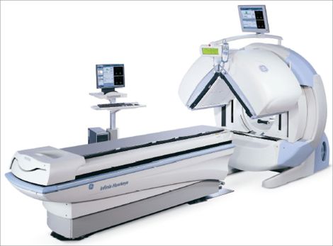
The Camera
For a MUGA scan, we acquire the images using a gamma camera. A gamma camera is a type of radiation detector that is used to show us how the radioactive labelled red blood cells are travelling through your left ventricle. Our cameras do not emit radiation or loud noises and are not a hazard to anyone in the vicinity while acquiring.
To acquire the images one of the camera heads above you will be brought in close to your left side at a 45-degree angle. The angle allows for the most optimal view of the left ventricle. This also means that above and to the right of you there will be no camera head.