- Unexplained bone pain
- Infection in the bone (osteomyelitis)
- Bone injury that can’t be seen on x-ray
- Arthritis
- Paget’s disease
- Cancer originating in the bone
- Cancer that has metastasized to the bone
- Impaired blood supply or death of bone tissue
Preparation
There is no required preparation for a bone scan; Eat, drink, and take your medications as normal.
PLEASE INFORM YOUR DOCTOR OR THE TECHNOLOGIST IF YOU
ARE, OR THINK YOU MIGHT BE, PREGNANT OR BREASTFEEDING.
Procedure
Check-in at the Diagnostic Imaging Department at the Sault Area Hospital for your appointment.
A bone scan is performed by injecting a tiny amount of a radioactive tracer (Technetium-99m Medronate) into a vein. An initial set of pictures may be obtained to evaluate the blood supply to the area of interest immediately following the injection.
You will then be asked to return to the nuclear medicine waiting room after sufficient time has elapsed to allow the radioactive tracer to localize in the bones and clear out the surrounding soft tissue. Typically it takes between 2 ½ to 4 hours for the injection to localize before we are able to take the delayed images.
During the wait, we require you to drink more fluids than normal and void your bladder frequently. A significant portion of the radioactive injection will be cleared out through your urine within the first 6 hours. To decrease radiation exposure to the pelvic area, we encourage frequent voiding. There are no other restrictions during this time.
When you return for the delayed images, you will be required to empty your bladder before imaging. A full bladder will degrade image quality in the pelvic region and may block visualization of pathology.
For the scan, you will be required to lie flat on the imaging bed while the camera slowly scans your bones. A whole-body scan typically takes less than 40 min; scanning a limited area will take less time. Additional views are sometimes required.
To better visualize some of your bones and help localize any pathology, we also acquire a Single Photon Emission Computerized Tomography (SPECT) image. SPECT imaging will create a three-dimensional model of your bones by slowly rotating the cameras around the area of interest. SPECT imaging usually takes between 12 and 15 minutes. When looking at the extremities (hands, feet, knees, etc.) or for infection, we may be required to do a SPECT/CT image of the area. A SPECT/CT image is similar to the SPECT image described above. However, a low dose CT image is acquired after the SPECT image, and the two images are fused together. A SPECT/CT image can further help the physician with a possible diagnosis.
Following your test, there are no restrictions, and you will be able to drive as you normally would. If you plan to go into the United States of America, you will set off the radiation detectors for approximately 2-3 days. We can provide a letter explaining why you are setting off the detectors; however, you will still be stopped.
Once your scan is complete, a radiologist will examine it, and a report will be sent to the ordering doctor within a week. Follow up your test with the ordering physician or your family doctor.
The Injection
The amount of radioactive tracer injected for a bone scan is small enough that there are no additional requirements or precautions to protect others from radiation exposure. The radiation exposure to you is a little more than a standard chest x-ray and less than half of that from a CT scan. Most of the injection will be cleared out of your body after 24 hours.
Allergic reactions to the material are extremely rare, and you should not feel anything or notice any changes from the material injected.
To get the injection to localize in your bones, we take advantage of your bones’ naturally occurring breakdown and remodelling. Like most other tissues in your body bones are constantly breaking down and remodelling themselves. As your bones remodel themselves they take in the radioactive tracer relative to the amount of remodelling. Bones that are remodelling more or repairing will take up more of the radiotracer. When we acquire your images, we can see areas of increased tracer uptake that represent areas of possible pathology.
The Camera
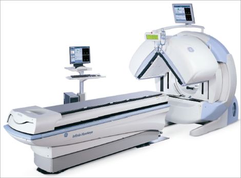
For a bone scan, we acquire the images using a gamma camera. A gamma camera is a type of radiation detector that is used to show us how the radioactive tracer is distributed throughout your bones. Our cameras do not emit any radiation unless performing a SPECT/CT and are not a hazard to anyone in the vicinity while acquiring.
Additionally, our cameras do not make any loud noises. The cameras we use have 2 detectors: one above you and one below you. Dual camera heads allow 2 views to be acquired simultaneously, and for SPECT imaging to be acquired in half the time.