A Lung Scan is a radiological procedure performed in the Nuclear Medicine department to evaluate the lungs. There are two parts to a lung scan: Ventilation and Perfusion. The ventilation part of the lung looks at air moving into and out of the lungs (bronchi and bronchioles). The perfusion part evaluates blood flow within the lungs.
Some reasons a lung scan may be ordered, but are not limited to:
- Evaluate a possible pulmonary embolism (if unable to have a CT with contrast)
- Functioning of different parts of the lung prior to lung surgery
- Identify areas of lung capable ventilation
Preparation
There is no required preparation for a lung scan; Eat, drink, and take your medications as normal.
If you have not had a chest x-ray in the 24 hours prior to a lung scan you will be required to have a chest x-ray which will be arranged for you at the time of your appointment.
PLEASE INFORM YOUR DOCTOR OR THE TECHNOLOGIST IF YOU ARE, OR THINK YOU MIGHT BE, PREGNANT OR BREASTFEEDING.
Procedure
Check-in at the Diagnostic Imaging Department at the Sault Area Hospital for your appointment.
A lung scan is typically performed in two back-to-back parts: ventilation; and perfusion.
For the Ventilation portion, you will be required to breathe in a radioactive aerosol (Technetium-99m Pentetate) for approximately 5 minutes through a tube with your nose plugged. If you are unable to breathe through the tube, a mask may be placed over the mouth and nose instead. Once enough of the aerosol has gotten into your lungs a SPECT (Single Positron Emission Computed Tomography) image will be taken under our camera. The images will show how gas (air) is being exchanged in your lungs when you breathe.
The second part of the test is the perfusion scan which will show how blood is travelling through your lungs. To perform the perfusion scan you will be given a small injection of a different radioactive tracer (Technetium-99m Macroaggregated Albumin [MAA]). The injection will travel to your lungs through your bloodstream and be taken into the lung tissue. Once the injection localizes in the lungs a second SPECT image will be acquired. The second set of images will demonstrate how blood is travelling through your lungs and will be acquired from the same angles as the ventilation scan.
The whole test typically takes around 40 minutes to complete.
Following your test, there are no restrictions and you will be able to drive as you normally would. If you are planning on going into the United States of America you will set off the radiation detectors for approximately 2-3 days. We can provide a letter explaining why you are setting off the detectors however you will still be stopped.
Once your scan is complete it will be examined by a radiologist and a report will be sent to the ordering doctor within a week. Follow up your test with the ordering physician or your family doctor.
The Injection
The amount of radioactive tracer used for a lung scan is small enough that there are no additional requirements or precautions to protect others from radiation exposure. The radiation exposure to you is less than a standard chest x-ray and far less than that from a CT scan. Most of the radioactive materials will be cleared out of your body after 24 hours.
Allergic reactions to the material are extremely rare, and you should not feel anything or notice any changes from the materials used.
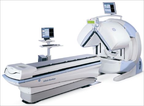
The Camera
For a lung scan, we acquire the images using a gamma camera. A gamma camera is a type of radiation detector that is used to show us how the radioactive tracers are distributed throughout your lungs. Our cameras do not emit radiation or loud noises and are not a hazard to anyone in the vicinity while acquiring. The cameras we use have 2 detectors: one above you and one below you. Dual camera heads allow for two views to be acquired at the same time decreasing the length of the test.